Introduction
Cardiovascular disease is the leading cause of death in the world. About a quarter of patients have no relevant symptoms before the onset of disease. Parameters obtained from cardiovascular imaging investigations, including coronary artery and aortic calcification, and epicardial fat volume, can provide important prognostic information and serve as risk-stratification tools. Our team is the pioneer in identifying that the epicardial fat amount and local thickness calculated by computed tomography (CT) images were closely related to aortic/coronary calcification, coronary plaque burden, and multiple cardiovascular risk factors in the late 2000s. To assess the amount of epicardial fat, researchers need to annotate the pericardium of all CT slices one by one, and it takes roughly 60 minutes for a case. Although non-contrast chest CT is widely used for early lung cancer screening, there is currently no tool to rapidly quantify cardiac/aortic calcification and epicardial adipose tissue.
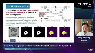
The National Taiwan University TW-CVAI (Taiwan-CardioVascular Artificial Intelligence) team jointly developed the "Automatic Pericardium/Aorta Segmentation AI Model (HeaortaNet)”. It is a one-stop analysis tool that integrates image segmentation, calculation of cardiovascular disease risk-related imaging parameters, and concatenated cardiovascular risk prediction models.
The HeaortaNet AI model is a deep learning model based on Unet, attention gate, and variational autoencoder/decoder structures. It had been trained by >70,000 axial images from 200 patients, with verified annotations of the pericardium and aorta. It shortens the time for data processing from 60 minutes, by manual segmentation of both pericardium and aorta, to 0.4 seconds. The segmentation accuracy is 94.8% for the pericardium, and 91.6% for the aorta.
The HeaortaNet technology has been applied patent in Taiwan and the United States (Taiwan Case No. 111120307; U.S. Case No. 17/804,839). It is the first officially approved AI model by National Taiwan University Hospital to assist detection and diagnosis in the Department of Medical Imaging in March 2021. At present, more than 5,000 non-contrast chest CT studies have been analyzed by the HeaortaNet. A dedicated cardiac imaging clinic facilitated by the HeaortaNet was opened at The National Taiwan University Hsin-Chu Branch in February 2022. The HeaortaNet 1.0 model was also validated by the Nvidia and placed at the Nvidia GPU Cloud open for global AI researchers.
The HeaortaNet 1.0 model had been applied to non-contrast chest CT images from more than 450,000 patients uploaded in the National Health Insurance Image Databank, proving its applicability across CT models. An imaging-based cardiovascular risk prediction model based on nationwide insurance claim-based data is under development. A web-based reminding system based on the analytic results of the HeaortaNet established in the National Health Insurance Administration to promote healthy behavior and early prevention for all people in Taiwan is about to be released. This will be the first of its kind nationwide implementation in the world.
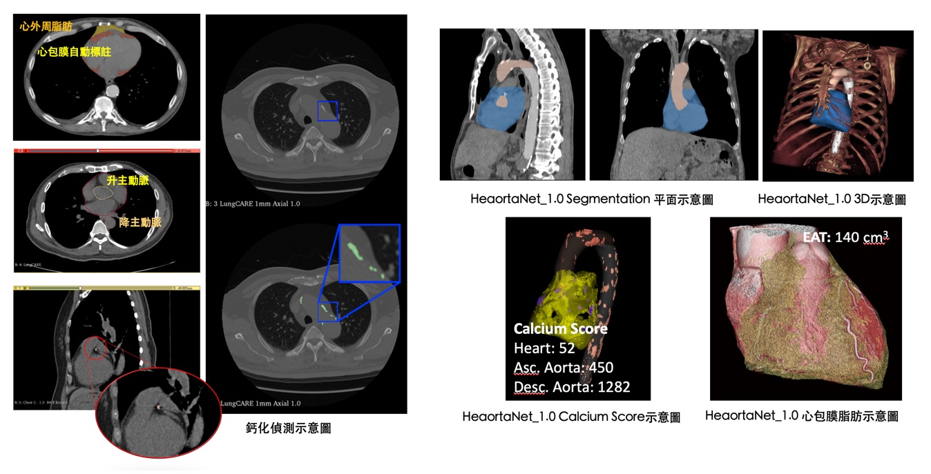
Our team keeps upgrading the HeaortaNet 1.0 model, so as to segment different sections of aorta and the heart, and to quantify calcification and adipose tissues along coronary arteries and aorta, respectively. The HeaortaNet was granted the 2021 Future Tech Award and the 18th Innovation Award.